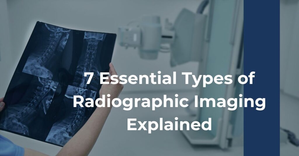
Radiographic imaging refers to the use of various techniques to Types of Radiographic create images of the inside of the body. These images are produced using different forms of radiation, such as X-rays or gamma rays, which can pass through the body and create a visual representation of the internal structures.
There are several types of radiographic imaging that are commonly used in healthcare:
Each type of radiographic imaging has its own advantages and limitations, and healthcare professionals may choose the most appropriate modality based on the specific condition being investigated. These advancements in technology have greatly improved our ability to diagnose and treat a wide range of medical conditions, ultimately enhancing the quality of care we receive.
1. Conventional X-Ray Radiography
When you think of radiographic imaging, the first picture that might pop into your mind is the conventional X-ray. It’s the quintessential diagnostic tool, familiar to anyone who has ever had a bone injury or a dental check-up. Despite being one of the oldest forms of medical imaging, it remains a cornerstone in the diagnostic process due to its simplicity, efficacy, and widespread availability.
- Simplicity and Speed: X-ray imaging is quick and easy to perform, which is why it’s often the first imaging technique used to assess various conditions.
- Visibility of Dense Structures: X-rays excel at providing sharp images of bones and can reveal fractures or dislocations clearly.
The anatomy illuminated by X-ray beams provides critical insights. A standard chest X-ray, for instance, can unveil the health of your lungs and heart. It’s also essential in tracking the progression of conditions such as osteoporosis or detecting foreign objects within the body.
There’s a fundamental reason why X-rays are a staple in the realm of types of radiographic imaging: they offer a straightforward glimpse at dense tissues, making them an indispensable part of acute care and preventative medicine.
2. Fluoroscopy
Imagine being able to watch a movie of your insides. That’s essentially what fluoroscopy offers. It’s a type of medical imaging that captures real-time moving body structures using a continuous X-ray beam.
- Real-Time Visualization: Fluoroscopy provides a dynamic view, which is particularly valuable during diagnostic procedures like barium swallows or catheter insertions.
- Interventional Use: Surgeons often rely on fluoroscopy as a navigational guide during procedures such as stent placements or joint replacements.
While traditional X-rays give a static snapshot, fluoroscopy is akin to giving your physician X-ray goggles, allowing them to see organs, like the heart and digestive tract, in motion. And when it comes to certain conditions or interventions, the real-time perspective is a game-changer. The continuous feed from fluoroscopy can be essentially transformative for both diagnostics and therapeutic interventions.
3. Computed Tomography (CT)
CT scans, a more intricate cousin of conventional X-rays, produce cross-sectional views of your body—a feat not possible with standard X-ray images. With the help of computer processing, multiple X-ray measurements from different angles are combined to create a three-dimensional picture.
- 3D Imaging: CT imaging provides a comprehensive view of the body’s internal structures, surpassing the two-dimensional limits of standard X-rays.
- Contrast Enhanced Detail: By using contrast materials, CT can illuminate particular areas, providing a clearer distinction between tissues.
Renowned for capturing every nook and cranny of the internal landscape, CT scans are invaluable when it comes to diagnosing internal injuries and conditions such as cancers or blood clots. This three-dimensional storytelling of your body’s interior can help your doctor make a more accurate diagnosis and develop a targeted treatment plan.
4. Magnetic Resonance Imaging (MRI)
If CT scans are storytellers, then MRIs are the poets of the imaging world. MRI uses powerful magnets and radio waves to generate detailed images of the body’s soft tissues, such as the brain, spinal cord, and joints.
- Soft Tissue Contrast: MRIs are particularly adept at differentiating between different types of soft tissue, making them essential in diagnosing a variety of conditions.
- No Radiation Exposure: Unlike X-rays and CT Scans, MRI doesn’t use ionizing radiation, which can be a significant advantage for patients requiring multiple scans.
This imaging technique is premier when the clarity of soft tissue is paramount—like in the intricate tapestry of the nervous system or the complex structure of joints. The level of detail it offers can provide a vital advantage in catching abnormalities early.
5. Digital Subtraction Angiography (DSA)
In the world of types of radiographic imaging focused on the body’s vascular system, Digital Subtraction Angiography stands out. DSA is a specialized form of fluoroscopy that provides a clear image of blood vessels by removing (subtracting) the surrounding bone and tissue densities from the pictures.
- Focused Vascular Imaging: DSA zones in on the body’s arterial network, searching for blockages or malformations.
- Enhanced Clarity: By subtracting unnecessary densities, DSA ensures vascular issues are not masked by other structures.
This imaging modality has revolutionized the way doctors approach procedures involving blood vessels. Blood flow can be watched in real time, and in the event of abnormalities—such as in the case of aneurysms or arterial blockages—the vascular narrative is crisply defined. It’s detailed enough to guide interventions, often helping to avoid more invasive surgical techniques.
6. Mammography
Mammography stands as a pivotal technique within the types of radiographic imaging, tailored specifically for breast tissue examination. This modality plays a vital role in early breast cancer detection, often capable of identifying tumors before they can be felt. Since early detection is key in the fight against breast cancer, mammography can truly be life-saving.
- Focused Breast Imaging: Mammography is the go-to method for visualizing breast tissue, adept at detecting abnormalities, including microcalcifications and tumors.
- Screening and Diagnostic Tool: It’s utilized as a preventive screening tool for asymptomatic women and as a diagnostic tool for those presenting symptoms.
Equipped with two primary types—screening mammography and diagnostic mammography—this specialized imaging form has distinct purposes. While the former is for routine check-ups, the latter zooms in on identified areas of concern, providing enriched detail where it matters most. The sophistication of mammography has evolved substantially, with innovations like 3D mammography offering enhanced detail and accuracy over traditional 2D images. This evolution in mammography techniques continues to advance the prospects of early cancer detection and effective treatment.
7. Dual-Energy X-Ray Absorptiometry (DEXA)
For those concerned with bone health, particularly in the context of osteoporosis, a Dual-Energy X-Ray Absorptiometry scan, or DEXA, provides key insights.
- Bone Density Measurement: DEXA scans rate your bone health by measuring BMD, using a minimal dose of ionizing radiation.
- Effective Monitoring Tool: They are also effective for tracking the progression of osteoporosis and the results of its treatment.
DEXA scans afford physicians a unique window into your skeletal strength, yielding information that can be pivotal for initiating treatments to strengthen bones or monitor existing conditions. The credible facts on DEXA’s abilities to assess fracture risk are invaluable to both patients and healthcare providers.
Radiation Dose and Safety
When discussing the types of radiographic imaging, it’s essential to weigh the importance of radiation dose and safety. The use of ionizing radiation in most radiographic imaging methods brings forth concerns about potential risks to health.
- Risk Minimization: Radiology departments adhere to the ‘As Low As Reasonably Achievable’ (ALARA) principle to minimize the risk of radiation exposure.
- Patient Protection: Technological advancements and safety protocols ensure the doses received are within safe limits and monitored closely.
Receiving a radiographic imaging exam means entrusting your health to trained professionals who are committed to employing doses of radiation that are safe and controlled.
Imaging Test Implementation
Behind every successful imaging test are the skilled professionals who perform and interpret these procedures. Understanding the implementation of these tests may ease any anxiety you may have and provide clarity on what to expect during your visit.
- Radiologists: Medical doctors specially trained to interpret the results of your imaging tests.
- Technique-Specific Training: Each modality requires specialized knowledge for optimal result acquisition and interpretation.
Whether you’re undergoing a CT scan, MRI, or any other type of radiographic imaging, the dedicated healthcare professionals involved play essential roles in both conducting the tests and interpreting the findings. When it comes to seeing inside the human body, these experts have the training not only to capture what’s beneath the surface but to decipher the stories those images tell.
Conclusion
Throughout this exploration of the different types of radiographic imaging, it’s clear that each modality offers unique benefits and capabilities. From the bone-focused imaging of DEXA scans to the dynamic, real-time views provided by fluoroscopy, these tools have revolutionized medical diagnostics. The detailed layers revealed by CT and MRI scans have unveiled nuances of the body’s inner workings, and mammography has heightened the fight against breast cancer with early detection.
As technology advances and our understanding of radiographic imaging deepens, we become increasingly equipped to tackle complex medical challenges. It’s a testament to the innovative spirit that propels the field of radiology forward. Whether you’re a patient, a healthcare provider, or simply an individual fascinated by medical technology, there’s a palpable sense of awe at the precision and capability afforded by these imaging types.

